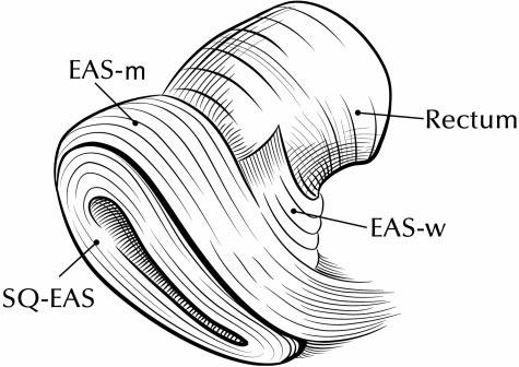
Fig. 2.

Drawing of external anal sphincter (EAS) subdivisions. Anterior portion of model is to the left, posterior to the right. Notice decussation of fibers toward the coccyx posteriorly. The main body of the external anal sphincter also has a concentric portion posteriorly that is not shown in this view. EAS-M, main body of EAS; EAS-W, winged portion of EAS; SQ-EAS, subcutaneous EAS.
