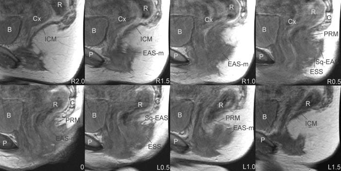
Fig. 5.

Sagittal magnetic resonance imaging images. Slices are ordered from right to left using the arcuate pubic ligament as reference. P, pubic bone; B, Bladder; Cx, cervix; R, rectum; ICM, ileococcygeus muscle; EAS, external anal sphincter; EAS-M, main body of external anal sphincter; PRM, puborectalis muscle; C, coccyx; ESS, external sphincter space; SQ-EAS, subcutaneous external anal sphincter; EAS, external anal sphincter.
