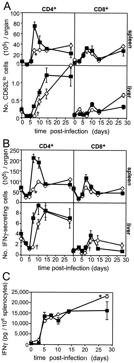
FIG. 3.
T-lymphocyte activation and IFN-γ secretion in spleens and livers of infected memTNF mice. WT (▪) and memTNF (⋄) mice were infected with 2,000 CFU of Listeria intravenously. (A and B) Single-cell suspensions were prepared from splenocytes and leukocytes from perfused livers of uninfected and infected mice at different times. Leukocytes were enumerated, stained for coexpression of CD4 or CD8 and surface CD62L (A) or intracellular IFN-γ (B), and detected using flow cytometry. (C) Splenocytes were cultured for 72 h with heat-killed L. monocytogenes or medium alone, and IFN-γ production was measured in the culture supernatant by an enzyme-linked immunosorbent assay. The data are the means and standard errors for five mice per group from one of two representative experiments. Significance for the spleen values was determined by ANOVA, and an asterisk indicates that the P value was <0.05 for a comparison of memTNF and WT mice. Significance for the liver values at day 7 was determined by Student's t test, and a dagger indicates that the P value was <0.03 for a comparison of memTNF and WT mice.

