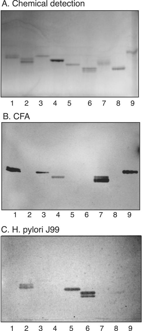
FIG. 3.
Comparison of glycosphingolipid recognition by CFA/I fimbriae of ETEC and BabA-expressing H. pylori. The glycosphingolipids were chromatographed on aluminum-backed silica gel plates and visualized with anisaldehyde (A). Duplicate chromatograms were incubated with 125I-labeled CFA/I fimbriae (B) and 35S-labeled H. pylori strain J99 (C), followed by autoradiography for 12 h, as described in Materials and Methods. The solvent system used was chloroform-methanol-water (60:35:8, by volume). Lanes: 1, Lea pentaglycosylceramide [Galβ3(Fucα4)GlcNAcβ3Galβ4Glcβ1Cer] (2 μg); 2, B type 1 hexaglycosylceramide [Galα3(Fucα2)Galβ3GlcNAcβ3Galβ4Glcβ1Cer] (2 μg); 3, Lex pentaglycosylceramide [Galβ4(Fucα3)GlcNAcβ3Galβ4 Glcβ1Cer] (2 μg); 4, B hepta-glycosylceramide (Galα3Galβ4GlcNAcβ3Galβ4GlcNAcβ3Galβ4Glcβ1Cer) (2 μg); 5, Leb hexaglycosylceramide [Fucα2Galβ3(Fucα4)GlcNAcβ3Galβ4Glcβ1Cer] (2 μg); 6, A type 1 heptaglycosylceramide [GalNAcα3(Fucα2)Galβ3(Fucα4)GlcNAcβ3Galβ4Glcβ1Cer] (2 μg); 7, Ley hexaglycosylceramide [Fucα2Galβ4(Fucα3)GlcNAcβ3Galβ4Glcβ1Cer] (2 μg); 8, A type 2 heptaglycosylceramide [GalNAcα3(Fucα2)Galβ4(Fucα3)GlcNAcβ3Galβ4Glcβ1Cer] (2 μg); 9, H type 2 pentaglycosylceramide (Fucα2Galβ4GlcNAcβ3Galβ4Glcβ1Cer) (2 μg). Autoradiography was performed for 12 h.

