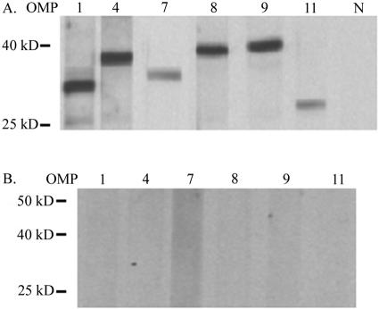
FIG. 3.
Expression of A. marginale OMPs in infected erythrocytes. (A) Western blots using A. marginale St. Maries strain-infected erythrocytes as an antigen. Polyclonal, monospecific mouse sera were used for detection of OMP1, OMP4, OMP8, OMP9, and OMP11, and monoclonal antibody 121/161 was used to detect OMP7. N, normal mouse serum. (B) Identical blots using uninfected erythrocytes with twice the protein load of panel A as a control. Predicted molecular sizes are as follows: OMP1, 32 kDa; OMP4, 37 kDa; OMP7, 39 kDa; OMP8, 40 kDa; OMP9, 40 kDa; OMP11, 32 kDa.

