
Figure 1.
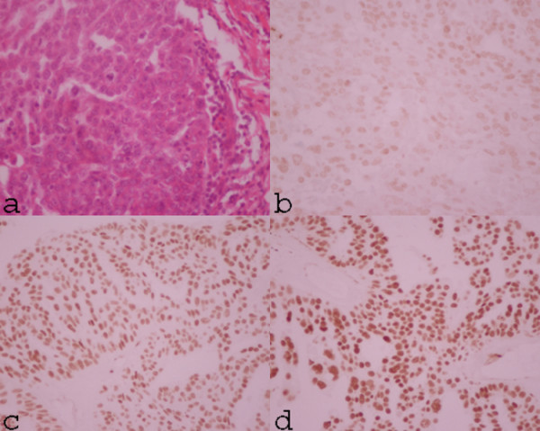
The H&E sections show a solid area of tumor which consists of cuboidal tumor cells with eosinophilic cytoplasm and vesicular nuclei, with prominent nucleoli. Mitotic figures are frequently observed (a). The immunohistochemical study of the same patient shows faint nuclei staining (intensity 1+) for WT1 protein in the most of tumor cells (b). The immunohistochemical studies show strong immunoreactivity in the nuclei in the majority of tumor cells (c: intensity 2+, d: intensity 3+)