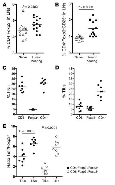
Figure 4. B16 melanoma TILs contain a high frequency of CD4+ and Foxp3+ but few CD8+ T cells.
The percentage of CD4+Foxp3+ T cells (A) and CD4+Foxp3+CD25– T cells (B) was analyzed by flow cytometry of lymph nodes from naive and tumor-bearing mice 15 days after tumor challenge. In a parallel set of studies, lymph nodes (C) and tumors (D) from tumor-bearing mice were analyzed at day 15 for their content of CD8+, Foxp3+, and CD4+ T cells. (E) The ratio of CD4+Foxp3– to Foxp3+ T cells (filled circles) and CD8+Foxp3– to Foxp3+ T cells (open circles) was calculated and compared in tumors and lymph nodes from tumor-bearing mice 15 days after tumor challenge. The data represent cumulative results from 3 independent experiments with 3–5 mice/group.

