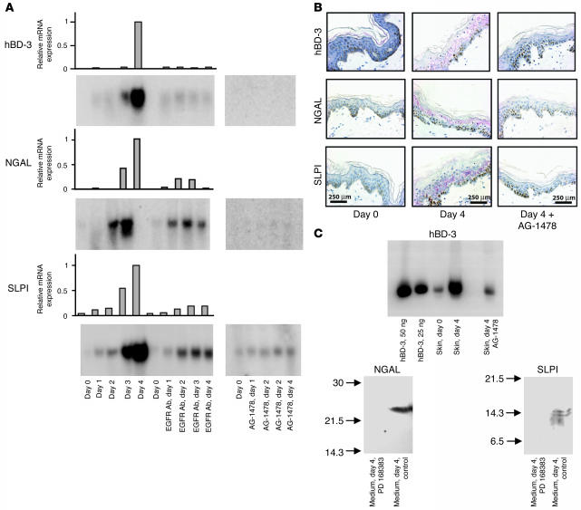
Figure 1. Expression of hBD-3, NGAL, and SLPI in wounded human skin.
Human skin was sliced into 1- × 10-mm slices and incubated for 0–4 days in culture. Each day, samples were processed for IHC or extracted for RNA or protein. (A) Northern blots of total RNA from whole skin. The blots were hybridized with probes for hBD-3, NGAL, SLPI, and G3PD. Graphs show the expression of hBD-3, NGAL, and SLPI normalized to the expression of G3PD mRNA. hBD-3, NGAL, and SLPI mRNA concentrations in the skin on day 4 were assigned the value 1. (B) Skin slices on days 0 and 4 with and without treatment with AG-1478 were stained for hBD-3, NGAL, and SLPI. Color was developed with fast red chromogen, and Harris hematoxylin was used for counterstaining. (C) The proteins extracted from both the wounded skin and the medium were analyzed by AU-PAGE and SDS-PAGE using synthetic hBD-3 as a standard and then blotted and probed with antibodies to hBD-3, NGAL, and SLPI. hBD-3 was only found in the skin. In contrast, NGAL and SLPI were only detected in the culture medium of the wounded skin. SLPI migrated as a double band around 14 kDa. This double band was not found in all samples tested and is probably due to proteolytic cleavage of SLPI during the preparation of this particular sample. AU-PAGE was used for detection of hBD-3 since this method is superior for examining small cationic peptides.

