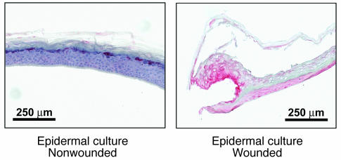
Figure 2. Expression of hBD-3 in wounded organotypic epidermal cultures.
Organotypic keratinocyte epidermal cultures were wounded by sterile incision with a scalpel. Four days after wounding, the wounded and nonwounded samples were immunostained for hBD-3. Color was developed with fast red chromogen, and Harris hematoxylin was used for counterstaining.

