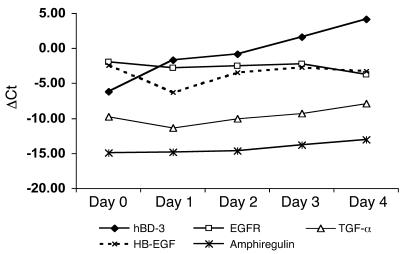
Figure 6. The expression of EGFR and EGFR ligands in wounded skin.
The expression of mRNA of EGFR, TGF-α, HB-EGF, amphiregulin, and hBD-3 was analyzed by real-time qRT-PCR. The mRNA expression is shown as the difference in threshold cycles between the gene of interest and G3PD. The expression of EGFR and its ligands was stable over time.

