
Figure 3. In vitro angiogenesis in IOMM-Lee parental and transfected cells.
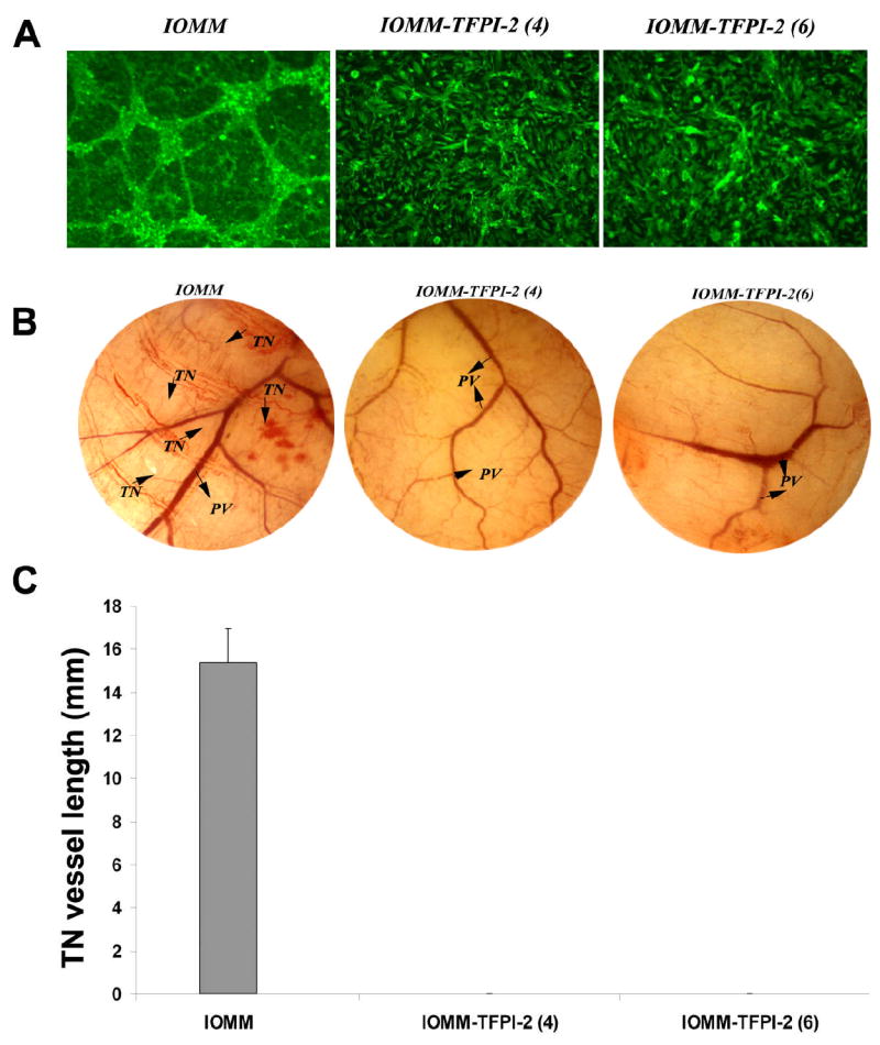
2×10⁴ IOMM-Lee parental and TFPI-2-transfected cells were plated on 8-well chamber slides and incubated overnight. 4×10⁴ human microvascular endothelial cells (HMEC) were added to the meningioma cells and co-cultured for 48 to 72 h. The co-cultures were then fixed and stained with factor VIII antibody and FITC-conjugated secondary antibody. The cells were observed under fluorescence microscopy for network alignment (A).2×106 IOMM-Lee parental and TFPI-2-transfected cells were suspended in 100–150 μL of serum-free medium, injected into the diffusion chamber, and the opening sealed and placed under the skin of nude mice for 10 days. The sc. implanted chamber resulted in microvessel formation (as indicated by arrows) with curved thin structures (TN) arising from pre-existing vessels in IOMM-Lee parental cells. In contrast, no angiogenesis was observed in TFPI-2-transfected cells (B). Quantification of the in vivo vasculature was done by measuring the length of the tumor induced vasculature (TN) (C).