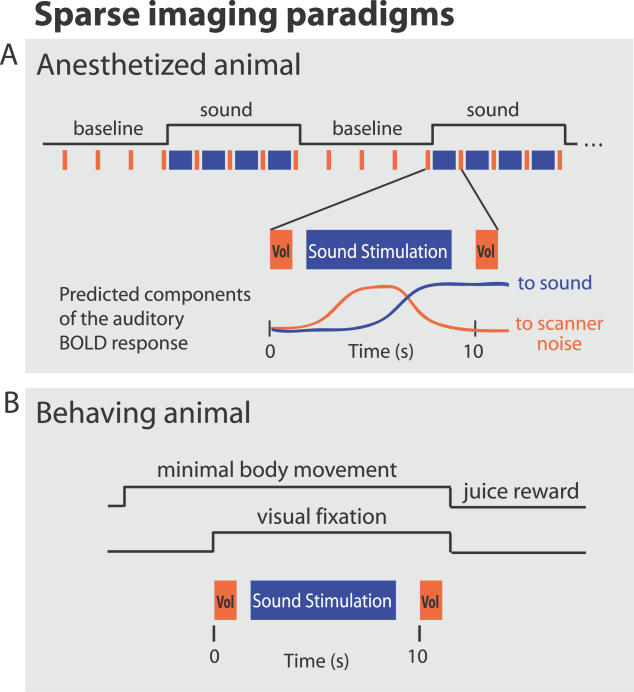
Figure 1. Sparse-Imaging Paradigms.
(A) The sparse-imaging paradigm for imaging the anesthetized animals (at 4.7- and 7-T) consisted of a block design with alternating stimulation and baseline periods. Data acquisition and sound stimulus were interleaved as schematized in the blow-out of a single sequence during a stimulation block. Here, the sequence initiates with an imaged brain volume, allowing sounds to be presented during the subsequent silent period. Because of the delay in the hemodynamic (BOLD) response the next volume acquired 10 s later reflects the sound stimulation that occurred approximately 6 s before. This minimizes influences on the auditory cortex BOLD response by the scanner noise that occurred 10 s before. This sequence is repeated four times during a stimulation block and four times during a baseline (no-stimulation) block.
(B) For imaging the behaving animal (at 7-T), the animal completed behavioral trials composed of the single sparse-imaging sequence during minimal eye and body movements (see “Behaving Animal Preparation and Imaging” in Materials and Methods).

