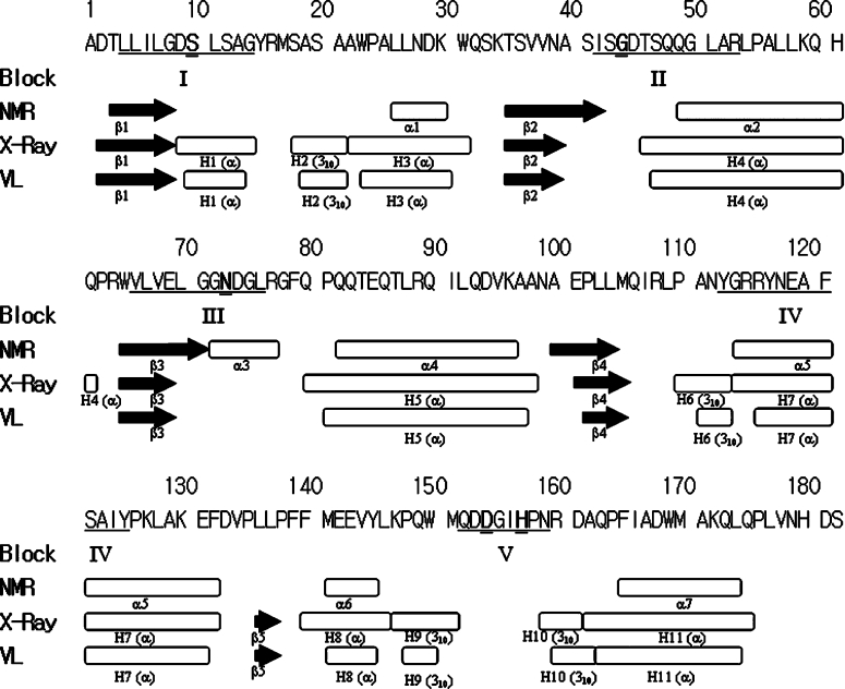
Figure 1. Secondary structures of TAP solved by NMR [19] and crystallography [22].
Black arrows represent β-strands and oblongs represent helices including α-helix and 310-helix. There are five sequence-conserved blocks (I–V) and the corresponding residues located in TAP are underlined. Ser10, Gly44, Asn73, Asp154 and His157 are marked in bold and are double-underlined. The structure solved by NMR is composed of four β-strands to form a four-stranded parallel β-sheet and seven α-helices. The structure solved by crystallography consists of a five-stranded parallel β-sheet, four 310 helices and seven α-helices (PDB code 1IVN). The co-ordinates of the crystal structure shown in the VL line and were plotted using the ViewerLite (version 5.0) program. This shows only minor differences compared with the crystal structure, which are the result of the slight differences in the secondary-structure definition.

