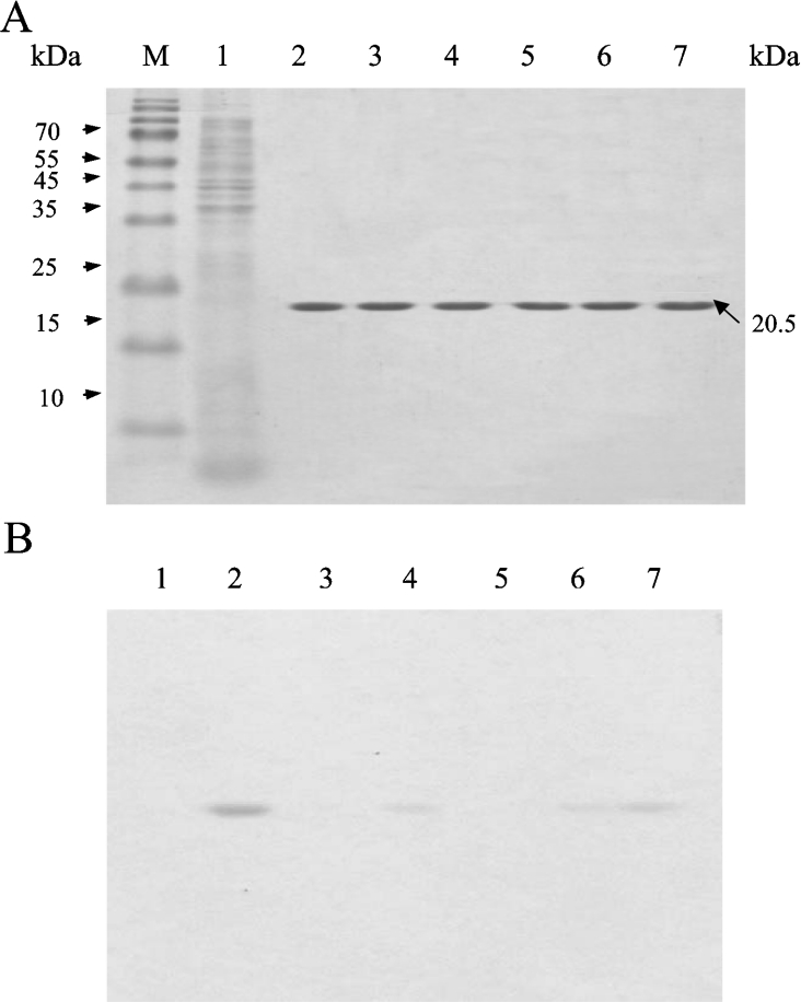
Figure 3. SDS/PAGE analysis of native and mutant enzymes using protein and activity staining.
Each protein (mutant and wild-type) was purified by affinity chromatography using Ni-NTA resin and was subjected to SDS/PAGE (15% gels, 1% SDS), with 2 μg of protein loaded into each well. (A) Coomassie Brilliant Blue-stained gel. (B) Esterase-activity staining (the assay was performed as described in the Experimental section). Lane M, molecular-mass markers (sizes given in kDa); lane 1, the crude extract of the host E. coli BL21(DE3) containing plasmid vector pET 20b(+) without TAP gene insertion; lane 2, wild-type TAP enzyme; lane 3, mutant S10A; lane 4, mutant D154A; lane 5, mutant H157A; lane 6, mutant G44A; lane 7, mutant N73A.

