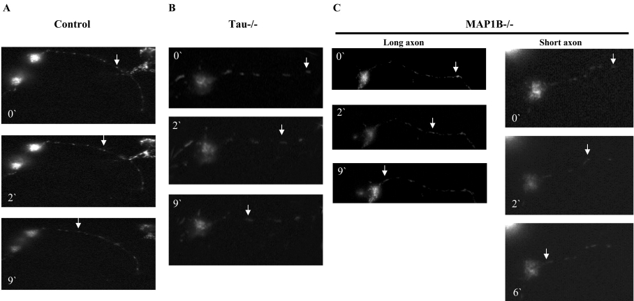
Figure 3. Retrograde mitochondrial transport in hippocampal neurons.
The movement of a single mitochondrion, at different times, is indicated for neurons from wild-type mice (A), tau−/− mice (B) and MAP1B−/− mice (C), in a long axon and in a short axon. Sample images from a video recording are shown. By looking at this video, it is possible to indicate that we were looking at the same (single) mitochondrion.

