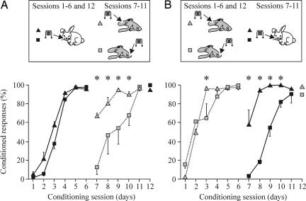
Fig. 3.
Acquisition of eyelid CRs during peripheral (vibrissae) followed by central (S1 areas for vibrissae or hind limb) CS, or vice versa. (A) Learning curves for groups 1 (black triangles) and 2 (black squares) in which the CS was presented during the first six sessions to the vibrissae and then (sessions 7–11) switched to the S1 area for vibrissae (group 1, gray triangles) or for the hind limb (group 2, gray squares). Data represent mean ± SD. Note that when the two central CSs were substituted by the peripheral CS, the acquisition was significantly faster for the group stimulated at the S1 area for vibrissae [∗, P < 0.001, F(11, 33) = 4.233]. (B) Learning curves for groups 3 (gray triangles) and 4 (gray squares) in which the CS was presented during the first six sessions to the S1 areas for vibrissae (group 3) or for the hind limb (group 4) and then (sessions 7–11) switched to the vibrissae (group 3, black triangles) or for the hind limb (group 4, black squares). Note that when the peripheral CS were substituted by the two central CSs, the acquisition was significantly faster for the group stimulated at the S1 area for vibrissae [∗, P < 0.05, F(11, 33) = 2.913].

