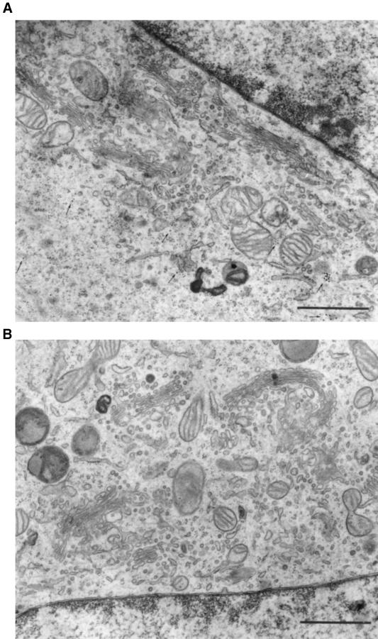
Figure 2.
Ultrastructure of NRK cells microinjected with N73pep. The cytoplasm of NRK cells was microinjected with the N73pep mixed with 10-nm protein A–gold as an injection marker (top; gold particles indicated by arrows). Cells were fixed after 1 h of incubation and processed for electron microscopy. In microinjected cells (top), the number of vesicles in the Golgi region was increased (see Table 1 for quantitations) compared with uninjected cells in the same section (bottom). Bar, 1 μm.

