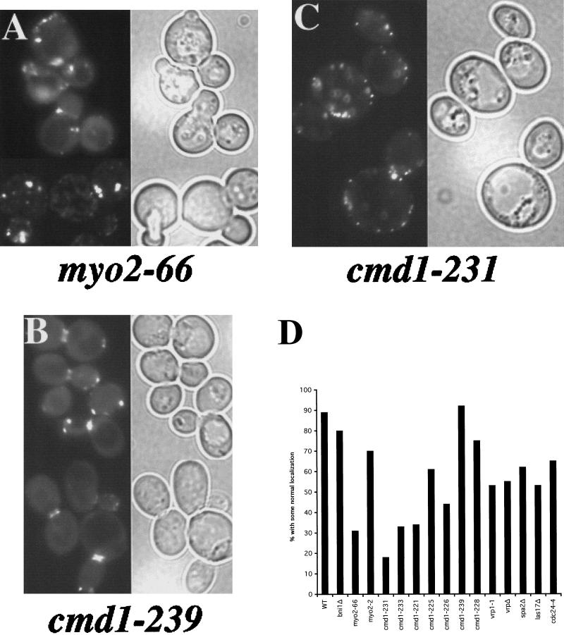
Figure 3.
The role of cell polarity proteins in Aip3p localization. The myo2-66 strain LWY2753 (A), the cmd1-239 strain DBY5719 (B), and the cmd1-231 strain DBY5711 carrying the GFP-Aip3p–expressing Cen vector pDAb204, were shifted to 37°C for 90 min and examined by fluorescence microscopy. GFP fluorescence is shown on the left of each panel, and a DIC view of the same cells is shown on the right of each panel. GFP-Aip3p localization was quantified in several cell polarity mutants after a shift to 37°C for 90 min (D). Individual cells were scored for the correct localization of some of their pool of GFP-Aip3p, and the values reported are derived from counts of 200–350 cells.

