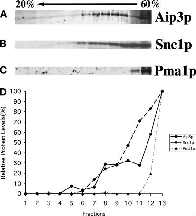
Figure 6.
Aip3p fractionates with a late secretory vesicle marker in a sucrose density gradient. The sec6-4 pep4Δ strain HJY3 was grown to 1 × 10⁷ cells/ml and shifted to 37°C for 2 h. Cells were collected and then spheroplasted and lysed in a Dounce homogenizer. The cell extract was clarified by centrifugation at 450 × g for 3 min, adjusted to 60% sucrose, underlaid in a 20–50% sucrose gradient, and centrifuged at 100,000 × g for 20 h. Thirteen fractions were collected from the top of the gradient, and samples from these fractions were separated on SDS-PAGE gels, transferred to nitrocellulose, and blotted with anti-Aip3p (A), anti-Snc1p (B), and anti- Pma1p (C) antibodies. Densitometry was used to quantify the relative amounts of protein in each fraction (D).

