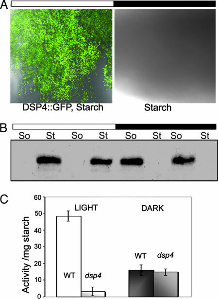
Fig. 5.
DSP4-GFP binding to starch in vivo. The amount of DSP4-GFP on the starch granules was detected by confocal microscopy (A) or GFP antibody (B). (A) The binding of DSP4-GFP to starch isolated in the day or night. Starch granules were isolated and observed under confocal microscope. Images were obtained by two subsequent scans with blue excitation light for GFP detection and DIC for starch visualization. The figure is typical of three independent experiments. (B) The amount of starch-bound DSP4-GFP (St) or DSP4-GFP in the soluble phase (So) was analyzed by Western blots. Samples at two sequential time points in the light period (8 and 14 h) and dark period (2 and 4 h) were analyzed. At each time point, 5 mg of starch or 150–200 μg of total protein from the soluble phase were subjected to SDS/PAGE and Western blotting. (C) Starch-bound phosphatase activity in the wild type and dsp4 after 14-h illumination in the day (Light) and after 2-h in the darkness (Dark). The mean values and standard deviations of two experiments for each data point are shown.

