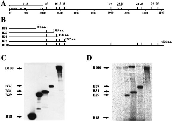
Figure 1.
Incorporation of [125I]iodopalmitate into secreted apoBs: apoB-29, apoB-31, apoB-37, and apoB-100 incorporate [125I]iodopalmitate, whereas apoB-18 does not. Distribution of cysteine residues in apoB-100 (A) and various apoB truncated constructs (B). Free cysteine residues are represented by long ticks, and short ticks represent cysteine residues found in disulfide linkages. (C) Western blot analysis of various secreted [125I]iodopalmitate-labeled apoB constructs with the use of the 1D1 anti-human apoB mouse mAb demonstrates the presence of the various apoBs on the PVDF membrane. (D) Incorporation of [125I]iodopalmitate into corresponding secreted apoB proteins visualized by autoradiography of the PVDF membrane. Exposure time was 3 d with the use of Molecular Dynamics phosphorimager cassettes.

