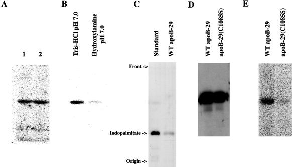
Figure 2.
ApoB-29 is palmitoylated on cysteine residue 1085 via a hydroxylamine-sensitive thioester bond. (A) Autoradiogram of [125I]iodopalmitate-labeled secreted WT apoB-29 starting material (lanes 1 and 2) blotted onto a PVDF membrane before hydrolytic treatment. (B) Autoradiogram of a PVDF membrane after a 72-h treatment with either 1 M Tris-HCl, pH 7.0, or 1.0 M hydroxylamine-HCl, pH 7.0. (C) TLC analysis of [125I]iodopalmitate standard and hydrolyzed radiolabel extracted from [125I]iodopalmitate-labeled apoB-29. Exposure time was 14 d on film with an intensifying screen. (D) Western blot analysis of WT apoB-29 and Cys1085Ser apoB-29 secreted from corresponding [125I]iodopalmitate-labeled McArdle-RH7777 stable cell lines. (E) Autoradiogram of WT apoB-29 and Cys1085Ser apoB-29 secreted from corresponding [125I]iodopalmitate-labeled McArdle-RH7777 stable cell lines. Exposure time was 3 d with the use of Molecular Dynamics phosphorimager cassettes.

