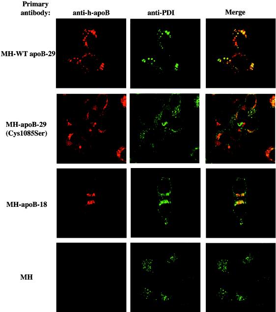
Figure 6.
Palmitoylation of apoB-29 is required for localization to a subcompartment of the ER. Colocalization studies of various apoBs with the ER marker protein PDI. Various human apoBs were detected with the 1D1 mouse mAb and are shown in red with the use of anti-mouse IgG-TR–conjugated secondary antibody (left panels). PDI was detected with the use of the rabbit polyclonal anti-PDI antibody and is shown in green with the use of anti-rabbit IgG-FITC–conjugated secondary antibody (middle panels). The merged images are shown in the right panels. Staining of McArdle-RH7777 cells stably expressing WT apoB-29, Cys1085Ser apoB-29, or apoB-18 and nontransfected McArdle-RH7777 cells is shown and identified as follows: MH-WT apoB-29, MH-apoB-29(Cys1085Ser), MH-apoB-18, and MH, respectively.

