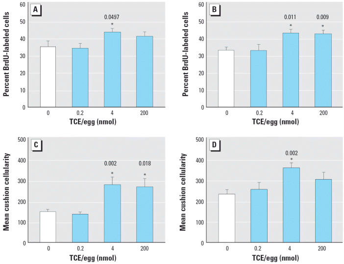
Figure 3.
Effects of TCE on cardiac cushion proliferation and cellularity in HH24 chick embryos. Embryos were exposed to TCE during cushion development. Percentage of BrdU-labeled cushion mesenchyme in (A) OFT cushions and (B) AVC cushions. Total mesenchymal cellularity in the (C) OFT cushions and (D) AVC cushions. Values shown are mean ± SE. Experiments were performed in triplicate; the total number of embryos evaluated was 8 for 0 nmol, 7 for 0.2 nmol, 10 for 4 nmol, and 9 for 200 nmol.
*Significantly different from 0 nmol treatment; p-values are given above bars.

