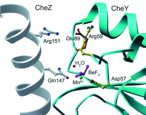
FIG. 2.
Active-site region of CheYN59R-BeF3−-Mn2+ structure modeled into the (CheY-BeF3−-Mg2+)2CheZ2 crystal structure (PDB code 1KMI). The secondary structures of CheY (cyan) and CheZ (gray) are in ribbon representation. Select residues in ball-and-stick representation are CheZ Gln147 (gray) and Arg151 (gray), CheY Asp57 and Arg59 (yellow), Glu89 (orange), BeF3− (magenta), and Mn2+ (green). A water molecule (red) has been modeled into the active site in the appropriate geometry for in-line attack of the BeF3− (analogue of PO32−) and within hydrogen-bonding distance of CheZ Gln 147 (29). Only one chain of the CheZ dimer is shown for clarity.

