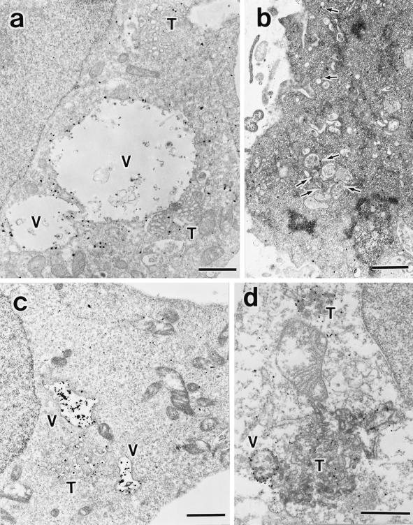
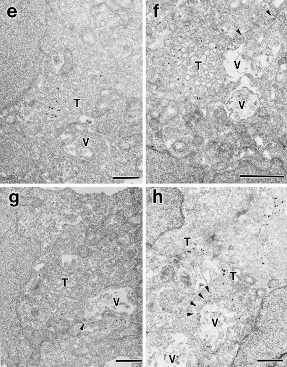
Figure 11.
Immunoelectron microscopy for GFP-SKD1E235Q, the endogenous SKD1, TfR, and EEA1. HEK293 cells transfected with GFP-SKD1E235Q were fixed, and the localization of GFP-SKD1E235Q (a), TfR (c), and EEA1 (e–h) was examined by silver-enhanced immunogold electron microscopy using antibodies against GFP, TfR, and EEA1, respectively. Distribution of the endogenous SKD1 in H-4-II-E cells was also examined using antibodies against SKD1 (b). (d) Double stain of GFP-SKD1E235Q (silver-enhanced gold particles) and TfR (HRP-DAB staining). V, exaggerated aberrant multivesicular vacuole; T, tubulo-vesicular structures. Arrows in b, endogenous SKD1 associated with membrane compartments/vesicles; arrowheads in f–h, EEA1 associated with protrusions of the vacuoles. Bar, 1 μm..


