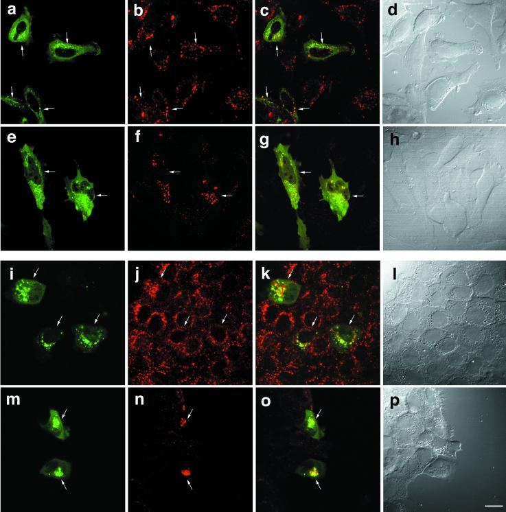
Figure 8.
Degradation of the internalized EGF is inhibited in the cells expressing GFP-SKD1E235Q. HeLa (a–h) and A-431 (i–p) cells transfected with GFP-SKD1E235Q were incubated at 4°C for 1 h in the presence of 3.3 and 0.1 μg/ml Texas Red-EGF, respectively. After washing, the cells were incubated at 37°C for 30 min (a–d and i–l) or for 3 h (e–h and m–p). Then the cells were fixed for fluorescence confocal microscopy. GFP-SKD1E235Q labeling (a, e, i, and m), Texas Red-EGF labeling (b, f, j, and n), a merged image (c, g, k, and o), and a differential interference contrast image (d, h, i, and p) are shown. Each row represents the same field. The cells expressing GFP-SKD1E235Q are indicated by arrows. Bar, 20 μm.

