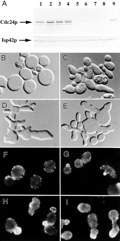
Figure 11.
Polarity establishment in the absence of Cdc24p. (A) Western-blot analysis using antibodies to Cdc24p and (as a control) the mitochondrial outer membrane protein Isp42p. (B–E) Cell morphologies as observed by DIC microscopy. (F–I) Immunolocalization of actin using anti-actin antibodies. Strain YEF1201 (cdc24Δ::HIS3 [pMGF5]) was transformed with plasmids YEp352-CDC42 and YEp13 (A, lanes 1 and 5, B, and F), YEp352-CDC42 and YEp13-CLA4* (A, lanes 2 and 6, C, and G), YEp352-CDC42 and YEp13-MSB1 (A, lanes 3 and 7, D, and H), or YEp352–42CLA4* and YEp13-MSB1 (A, lanes 4 and 8, E, I). Cells were grown to exponential phase at 30°C under mildly inducing conditions for GAL1-CDC24 (SC-Leu-Ura liquid medium containing 2% glucose plus 2% galactose), and samples were removed for immunoblot analysis (A, lanes 1–4). Cells were then shifted to repressing conditions (SC-Leu-Ura liquid medium containing 2% glucose only) for 12 h, diluted further with the same medium, and incubated for an additional 4 h before harvesting for immunoblot (A, lanes 5–8) and microscopic (B–I) analysis. Wild-type strain YEF473 growing exponentially in liquid SC medium at 30°C was used as a control (A, lane 9). B–I are printed at the same magnification.

