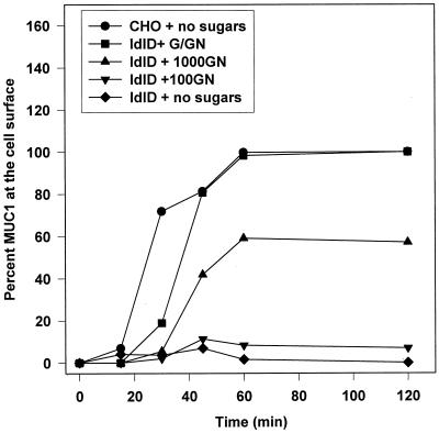
Figure 3.
MUC1 with truncated O-glycans reaches the cell surface with normal kinetics. Both CHO and ldlD cells expressing MUC1 were pulsed with [35S]Met/Cys for 15 min and chased for the times indicated before biotinylation of the cell surface. Varying levels of Gal and GalNAc were included in the starvation, pulse, and chase media as indicated (100 μM GalNAc, +100GN; 1000 μM GalNAc, +1000GN; 1000 μM GalNAc and 100 μM Gal, +G/GN). Biotinylated [35S]MUC1 was recovered with avidin-conjugated beads from the immunoprecipitates and subjected to SDS-PAGE for analysis of radioactive bands with a Bio-Rad phosphoimager system. Cell surface MUC1 levels in ldlD samples were normalized to the maximal level of cell surface [35S]MUC1 synthesized in ldlD cells in the presence of both Gal and GalNAc (+G/GN). The absolute levels of MUC1 expression in CHO and ldlD cells (+G/GN) are comparable (see Figure 5B).

