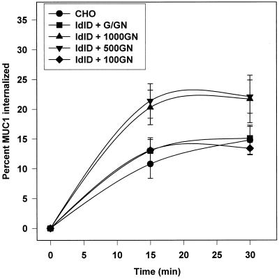
Figure 4.
MUC1 internalization from the cell surface is affected by its glycosylation state. CHO and ldlD cells expressing MUC1 were pulse labeled for 30 min and chased for 90 min before cell surface biotinylation on ice as described in MATERIALS AND METHODS. Varying levels of Gal (G, 100 μM) and GalNAc (GN, 100, 500, or 1000 μM) were included in the starvation, pulse and chase media as indicated. Cells were rapidly warmed to 37°C for the indicated times to allow internalization of surface MUC1 and rapidly cooled on ice, and remaining cell surface biotin was stripped with MESNA. Some samples were not treated with MESNA in order to determine the total amount of biotinylated MUC1 at t = 0. Internalized biotinylated MUC1 was recovered from the MUC1 immunoprecipitates with avidin-conjugated beads, and [35S]MUC1 was analyzed after SDS-PAGE using a phosphoimager. Data are plotted as the percent of total biotinylated [35S]MUC1 internalized at each time point and are presented as means ± SD of triplicate samples. Similar results were obtained in six experiments.

