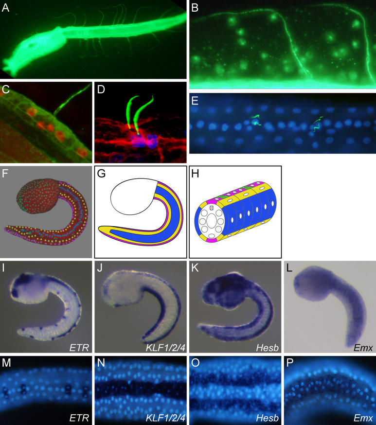
Figure 1. CESNs and Tail Epidermis Medio-Lateral Patterning.
(A–C) β-tubulin immunostaining showing CESN projections in the fin tunic. Confocal images projection with nuclei in red (propidium iodide) (C).
(D and E) Acetylated α-tubulin showing the proximal part of the projections from paired CESNs (Acetylated α-tubulin in green, DAPI in blue, phalloidin in red).
(F) 3-D projection extracted from a time lapse movie of an embryo injected with H2B-YFP mRNA and Fast Green (red). Trunk nuclei are green, while tail nuclei are colored to highlight the longitudinal rows made of single cell.
(G and H) Schematic representation of the tail medio-lateral patterning (medial row, purple; medio-lateral row, yellow; lateral row, blue).
(I–P) Expression patterns of tail epidermis markers at the mid-tailbud stage. Lateral view (I–L and P). Ventral view (M–O). Anterior is to the left. DAPI staining of nuclei in (M–P).

