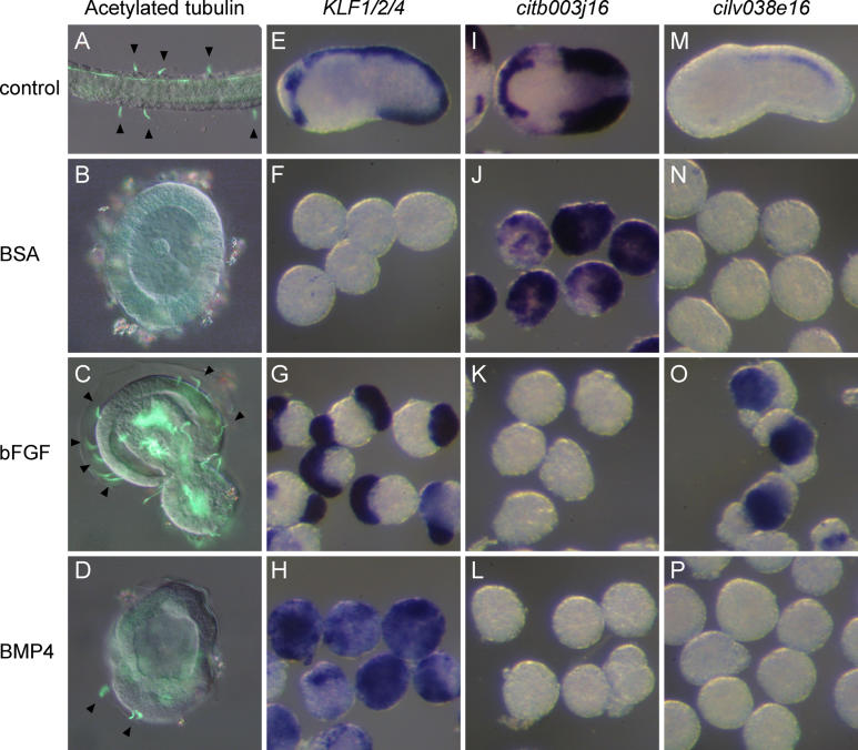
Figure 3. bFGF and BMP4 Induce Midline Fate and ESN Formation in Isolated b-Line Explants.
b4.2 Blastomeres were isolated at the eight-cell stage and treated with proteins from the 16-cell stage.
(A–D) Acetylated α-tubulin immunostaining at larval stage: CNS and PNS structures are labelled. Arrowheads indicate ESNs.
(E–P) Molecular marker expression at early tailbud stages: midline marker KLF1/2/4 (E–H), lateral and medio-lateral marker citb003j16 (I–L), and tail nerve cord marker cilv038e16 (M–P).
Top row, whole embryos (A,E,M) lateral view, (I) dorsal view, anterior to the left.
Second row, control b4.2 blastomeres treated with BSA.
Third row, b4.2 blastomeres treated with bFGF protein.
Bottom row, b4.2 blastomeres treated with BMP4 protein.

