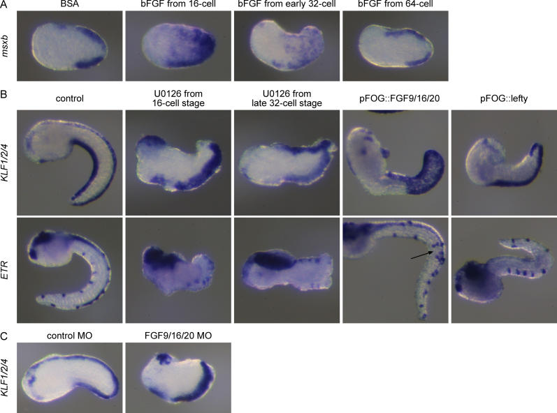
Figure 4. The FGF and Nodal Pathways Control Dorsal Midline and CESN Formation.
(A) bFGF-treated embryos show ectopic expression of the midline marker msxb at the late neurula stage when the treatment starts at the 16-cell or early 32-cell stages. Embryos do not respond to bFGF at the 64-cell stage.
(B) Blocking Erk activity with the pharmacological inhibitor U0126 from the 16-cell stage abolishes dorsal KLF1/2/4 and ETR expression at tailbud stages, while treatment from the late 32-cell stage has no effect on these markers. FGF9/16/20 is sufficient to induce ectopic midline and ESNs formation (arrow), while Lefty prevents dorsal midline formation.
(C) FGF9/16/20 MO–injected embryos show a loss of dorsal KLF1/2/4 expression while embryos injected with a control MO are not affected. (Lateral view, anterior to the left, dorsal to the top in all panels).

