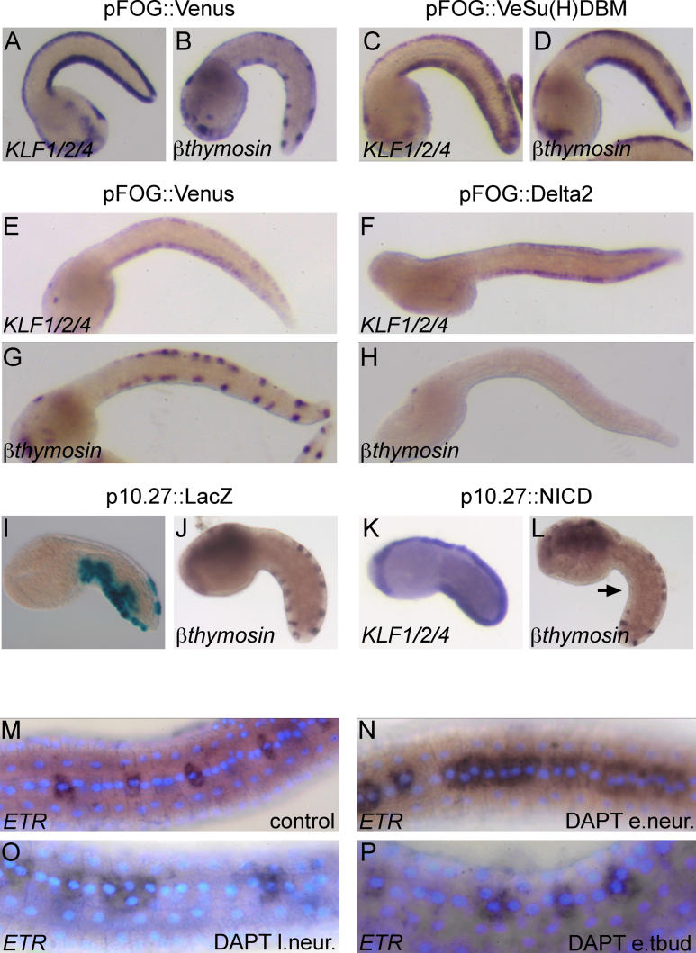
Figure 6. Delta/Notch Signalling Controls the Number of CESNs within Neurogenic Midlines.
(A,B,E,G) Electroporation of the construct pFOG::Venus (a bright YFP) has no effect on the midline domain and the CESNs, respectively, revealed by the KLF1/2/4 and β-thymosin-like probes. The expression levels of KLF1/2/4 at mid- to late tailbud stages (E) are lower than at mid-tailbud stages (A).
(C and D) Electroporation of the dominant-negative construct pFOG::Venus-Su(H)DBM does not affect the expression of KLF1/2/4 but leads to an increase in the number of β-thymosin-like-positive cells.
(F and H) Electroporation of pFOG::Delta2 results in a loss of β-thymosin-like-positive cells, without affecting KLF1/2/4 expression.
(I and J) The construct p10.27::LacZ preferentially drives expression of β-galactosidase in the ventral tail epidermis (I), without affecting the development of CESNs (J).
(K and L) Electroporation of p10.27::NICD, carrying an active form of Notch, does not affect the formation of the midline domain (K), but leads to a loss of CESNs in the ventral midline (arrow in L).
(M–P) Treatment with the γ-secretase inhibitor, DAPT, at various developmental stages results in varying degrees of midline epidermal cell replacement by ETR-positive CESN precursors. e.neur., early neurula; l.neur., late neurula; and e.tbud, early tailbud.

