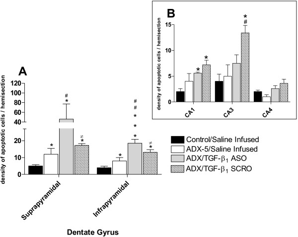
Figure 5.

Quantitative analysis of the effect of TGF-β1 ASO and SRO administration into the left lateral ventricle on hippocampal apoptotic morphology 5 days after adrenalectomy (ADX). Data represent the density of apoptotic cells (mean ± SEM) in the hippocampus ipsilateral to the side infused with the oligodeoxynucleotide or a saline solution. (A) Apoptosis observed in the suprapyramidal and infrapyramidal of dentate gyrus. Black columns, saline infused sham-operated animals; white columns, saline infused, ADX-5 animals; gray columns TGF-β1 ASO infused ADX-5 animals; dashed columns, TGF-β1 SCR infused ADX-5 animals. * p < 0.05 vs control; *** p < 0.01 vs control; # p < 0.05 vs ADX-5; ## p < 0.01 vs ADX-5, ≠ p < 0.05 vs ADX/TGF-β1 ASO. (B) Inset representing Cornus Amonis areas. Black columns, saline infused, sham-operated animals; white columns, saline infused, ADX-5 animals; gray columns TGF-β1 ASO infused ADX-5 animals; dashed columns, TGF-β1 SCRO infused ADX-5 animals. * p < 0.05 vs control; # p < 0.05 vs ADX-5.
