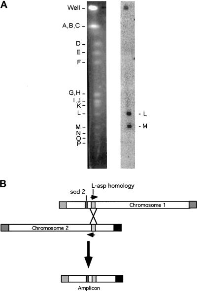
Figure 7.
Hybridization of the 180-kb novel joint region with NotI fragments L and M. (A) Wild-type S. pombe DNA, digested with NotI, was separated by pulsed-field gel electrophoresis. Left, ethidium bromide–stained gel. Right, hybridization with novel joint region fragment. The 320-bp novel joint region fragment was an EcoRV fragment of plasmid 1.1D. The experiment was repeated with a smaller probe from the centromere-proximal sequence of the novel joint, which hybridized only to NotI fragment M (our unpublished results). (B) A homologous recombination model for the 180-kb amplicon. The L-asp homologue on chromosome I is oriented with the direction of transcription away from the telomere (our unpublished results). sod2 is represented as a dark gray rectangle. The L-asp homology region is represented as a light gray rectangle. The arrows denote the direction of transcription of the L-asp homologues. The squares represent the telomeres: the chromosome I long arm telomere is gray and the chromosome II short arm telomere is black. The horizontally striped box is the telomere of the short arm of chromosome I, and the vertically striped box is the telomere of the long arm of chromosome II.

