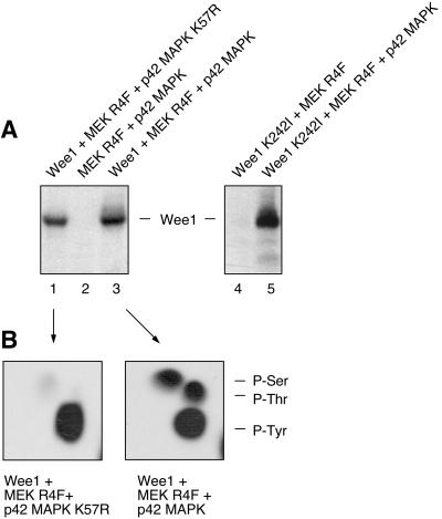
Figure 2.
Phosphorylation of Wee1 by p42 MAPK in vitro. (A) Autoradiogram of an in vitro phosphorylation reaction with purified, recombinant proteins and [γ-32P]ATP. Wild-type Wee1 was used in the experiment shown on the left; it exhibited substantial autophosphorylation (lane 1), with MEK R4F and p42 MAPK causing additional phosphorylation (lane 3). Kinase-minus Wee1 (Wee1 K242I) was used in the experiment shown on the right. It was phosphorylated in the presence of MEK R4F and p42 MAPK (lane 5), not in the presence of MEK R4F alone (lane 4). (B) Phosphoamino acid analysis of excised Wee1 bands from lanes 1 and 3 of A. Positions of ninhydrin-stained phosphoamino acid standards are indicated on the right.

