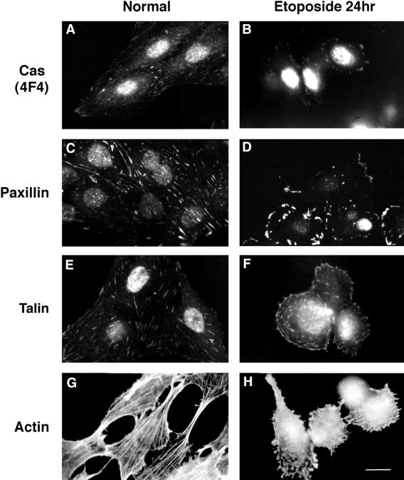
Figure 9.
Fluorescent images depicting the changes in the cellular localization of Cas, paxillin, talin, and actin in apoptotic cells. The distribution of Cas (4F4, A and B), paxillin (C and D), and talin (E and F) within control cells or etoposide-treated cells reveals that Cas, paxillin, and talin are localized in focal adhesion sites in control cells (A, C, and E) but is lost from focal adhesions during apoptosis and redistributes into the periphery of cells (B, D, and F). Actin labeling using TRITC-phalloidin reveals that the actin stress fibers seen in control cells (G) are virtually absent from the cytoplasm of apoptotic cells, although some truncated fibers are seen at the cell margins (H). Bar, 10 μm.

