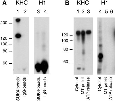
Figure 5.
H1 does not recognize conventional KHC in rat liver Golgi membranes or in Xenopus egg cytosol. (A) Ubiquitous kinesin immunoprecipitated from solubilized rat liver Golgi membranes using the SUK 4 monoclonal (lanes 1 and 3) was immunoblotted with either polyclonal anti-uKHC (lane 1), which recognized bands at ∼125 and ∼250 kDa (probably incompletely dissociated kinesin heavy chains), or H1 (lane 3), which failed to produce a signal. Nonimmune mouse IgG was used as a control (lanes 2 and 4). The bands between 50 and 60 kDa are antibody heavy chains (lanes 3 and 4). (B) Microtubule pellets enriched in motors were prepared from interphase Xenopus egg cytosol and then extracted with 5 mM ATP and 100 mM NaCl to give an ATP release fraction (lanes 3 and 6) and the remaining microtubule pellet (lanes 2 and 5). Interphase cytosol is loaded in lanes 1 and 4. After SDS-PAGE, protein samples were immunoblotted with either the polyclonal anti-uKHC antibody (lanes 1–3) or the H1 monoclonal antibody (lanes 4–6). Although H1 recognized a band at ∼70 kDa (lane 4), this polypeptide did not pellet with microtubules, unlike uKHC (compare lanes 2 and 3 with 5 and 6).

