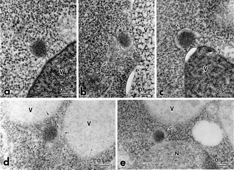
Figure 8.
prAPI is membrane associated in the apg5Δ mutant. The apg5Δ strain was grown to log phase in YPD and shifted to SD(−N) medium for 3 h to induce autophagy. Samples were prepared for electron microscopy with the use of rapid freezing and freeze-substitution fixation and stained with lead citrate for 1 min (a–c) or stained with lead citrate for 30 s and immunostained with anti-API antibodies (d and e), as described in MATERIALS AND METHODS. The arrows mark membranous structures around the Cvt complex. V, vacuole; N, nucleus.

