
Figure 2.
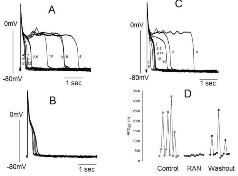
Ranolazine (RAN) reduces APD variability in left ventricular myocytes isolated from canine failing hearts. Twelve consecutive APs recorded at a pacing rate of 0.25 Hz are superimposed. Panel A: APs in the absence of drugs (control). Panel B: APs recorded in the presence of 10 μM of ranolazine. Panel C: APs recorded 3–4 minutes after drug washout. Panel D: Summary of the data shown in panels A-C. Each data point represents the APD50 from a series of APs recorded consecutively at a pacing rate of 0.25 Hz. The numbers next to the AP tracings in panels A and C are consecutive APs recorded during a period of ~ 45 seconds.