
Figure 4.
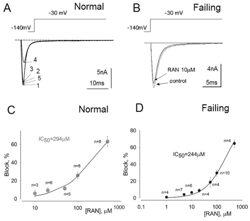
Ranolazine (RAN) weakly inhibits peak transient sodium current.
Effect of ranolazine on peak INa recorded from left ventricular myocytes isolated from normal (A, C) and failing canine hearts (B,D). Panel A: Superimposed current traces recorded in a non failing myocyte in the absence of drugs (1), and in the presence of 10 (2), 20 (3) and 500 μM ranolazine (4) and after drug washout (5). Panel B: Superimposed current traces recorded without (control) and with 10 μM ranolazine recorded in failing myocytes. Panels C and D: Concentration-response relationships for inhibition of peak INa by ranolazine in normal (C) and failing hearts (D). Points and error bars represent mean and standard error of peak sodium currents obtained in 13 myocytes from 2 normal (non-failing) hearts (C) and 14 myocytes from 4 failing hearts (D). Data points are fitted to a one-to-one binding model (solid line; Equation 1, Methods). The IC50 value is the concentration of ranolazine that caused 50% inhibition of peak INa.