
Figure 2.
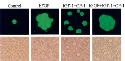
Chondrocyte appearance and pericellular matrix production after alginate culture in the presence of bFGF, IGF-1, and OP-1. Chondrocytes were cultured for 21 days in alginate beads in serum-free mini-ITS+ medium (control) or the control medium supplemented with 50 ng/ml bFGF, 100 ng/ml each of IGF-1 and OP-1, or the combination of all 3. At the end of the culture period, the beads were incubated with Calcein AM and ethidium bromide homodimer, as described in Materials and Methods, to assess survival. The beads were dissolved in sodium citrate, and a sample of the cells from each condition was observed using a fluorescence microscope (top). Dissolved alginate beads from separate cultures were pelleted by centrifugation, resuspended in Dulbecco’s modified Eagle’s medium, and placed in the bottom of microwell plates, followed by the addition of fixed erythrocytes, as described in Materials and Methods. A representative sample was photographed using an inverted phase-contrast microscope (bottom). The cell-associated (pericellular) matrix can be seen excluding the erythrocytes from the chondrocyte plasma membrane in the cells treated with IGF-1 plus OP-1. The clusters of cells treated with bFGF revealed by Calcein AM staining were partially broken up by the processing for the particle exclusion assay. See Figure 1 for definitions. (Original magnification × 400.)