
Figure 6.
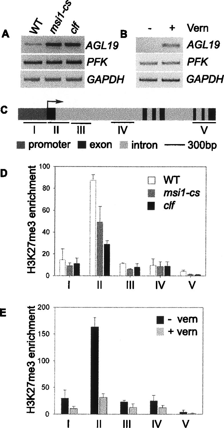
Histone methylation pattern at the AGL19 locus. (A) Expression analysis of AGL19 in msi1-cs and clf in rosette leaves of 4-wk-old plants grown in LD. (B) Expression analysis of AGL19 in 10-d-old seedlings grown in SD with or without a 6-wk vernalization treatment (Vern). (C) Schematic representation of AGL19 showing intron–exon structure (annotated according to TIGRv5) and position of the fragments used for amplification by PCR following ChIP. The arrow indicates the transcription start site. (D) Quantification of multiplex PCR products after ChIP using anti-trimethyl-histone H3K27 (H3K27me3) antiserum on 4-wk-old rosette leaves from wild-type (WT), msi1-cs, and clf plants grown in LD. Values are shown as enrichment of H3K27me3 using AGL19 specific primers relative to PFK for wild-type (WT), msi1-cs, and clf plants. (E) ChIP experiment performed on 10-d-old wild-type (WT) seedlings grown in SD with or without a 6-wk vernalization treatment using antibodies against H3K27me3. Values represent mean ± range from two independent ChIP experiments.