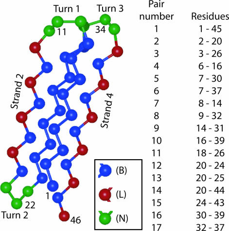
Fig. 1.
Protein model. (Left) A cartoon of the four-stranded β-barrel model used in this study. Each amino acid is represented by a single bead of diameter σ. The tether points, residues 1, 11, 22, 34, and 46, are on the outside of the native structure of the protein and are labeled, along with strands 2 and 4. (Right) A list of the representative contact pairs used to examine the folding mechanism through Pform. A list of native contacts for this protein is given in Table 2, which is published as supporting information on the PNAS web site.

