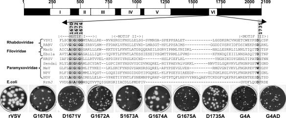
Fig. 1.
AdoMet-binding site alterations. (Upper) Amino acid sequence alignments of a predicted AdoMet-binding region of domain VI of nsNS RNA virus L proteins and known RNA methylases. The conserved motifs of nsNS RNA virus polymerases (I–VI) are shown (28). The AdoMet-binding residues modified in this study are shaded. VSVI, VSV Indiana; RABV, rabies virus; Marb, Marburg; HRSV, human respiratory syncytial virus; MeV, measles virus; NPV, Nipah virus; NDV, Newcastle disease virus; RrmJ, Escherichia coli 2′-O MTase. (Lower) Plaque morphology of recombinant viruses on Vero cells. Plaques of rVSV and G1674A were developed after 24 h; those of G1670A, G1672A, G1675A, D1735A, D1671V, and S1673A were developed after 48 h; those of G4A and G4AD were developed after 96 h.

