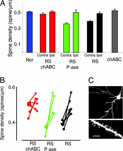
Fig. 4.
chABC combined with RS causes a significant recovery of dendritic spine density. (A) Mean spine density in normal rats (Nor), RS rats, RS P-ase-treated rats (RS P-ase), RS chABC-treated rats (RS chABC), and nondeprived rats treated with chABC (chABC). The data of RS, RS P-ase, and RS chABC are reported separately for the cortex ipsilateral (Ipsi) to the long-term-deprived eye that is untreated and the cortex contralateral (Contra) to the long-term-deprived eye that is treated with chABC or P-ase. In both RS P-ase and RS rats, spine density in the cortex contralateral to the deprived eye is smaller than in normal rats. In contrast, spine density in RS chABC-treated rats did not differ from normal rats (one-way ANOVA, P < 0.001; post hoc Tukey test, Nor vs. RS and RS P-ase, P < 0.001; Nor vs. RS chABC, P = 0.4). Spine density in the cortex ipsilateral to the deprived eye was not different among groups (Nor, RS, RS P-ase, RS chABC; one-way ANOVA, P = 0.82). The chABC treatment per se does not affect spine density (chABC vs. Nor, P = 0.42, t test). (B) Within-animal comparison of spine density in RS, RS P-ase, and RS chABC. Data from single animals are reported. Filled circles represent data for the cortex contralateral to the long-term-deprived eye; open circles represent values for the cortex ipsilateral to the long-term-deprived eye. Data from the same animal are connected by a segment. In RS and RS P-ase, density is significantly smaller in the contralateral cortex (RS P-ase, P = 0.037; RS, P = 0.003; paired t test). No difference is present between the two cortices of RS chABC rats (P = 0.29, paired t test). (C) Example of a layer II–III pyramid cell of the binocular visual cortex and of a dendritic branch. (Scale bar in C: Upper, 40 μm; Lower, 5 μm.)

