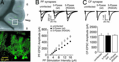
Fig. 1.
In vivo expression of IP3 5-Ppase in PCs decreases the PF–EPSC. (A) (Upper) Low-magnification image of cerebellar slice injected with virus carrying gene encoding IP3 5-Ppase (wt) and EGFP in vivo. Arrowhead indicates the site of the insertion of the viral injection pipette. (Lower) High-magnification image of region in white rectangle in Upper. GFP-expressing PCs were clearly imaged. (B) Representative traces of PF–EPSCs for indicated conditions of increasing stimulus intensities of 2, 8, 12, and 16 μA (holding potential = −80 mV). The amplitudes of PF–EPSC in uninfected PCs (○, n = 12), IP3 5-Ppase (wt)-expressing PCs (●, n = 11), and IP3 5-Ppase (R343A)-expressing PCs (◇, n = 12) are plotted as a function of stimulus intensity, indicating a decreased synaptic efficacy at PF synapses by in vivo expression of IP3 5-Ppase in PCs. (C) Representative traces of CF–EPSC for indicated conditions (holding potential = −10 mV). The amplitudes of CF–EPSC recorded from uninfected PCs (n = 10), IP3 5-Ppase (wt)-expressing PCs (n = 11), and IP3 5-Ppase (R343A)-expressing PCs (n = 11) indicate no significant decrease in CF–EPSC.

