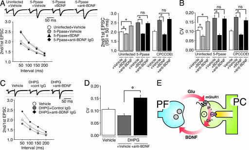
Fig. 5.
Application of BDNF or anti-BDNF IgG simultaneously with modulation of postsynaptic IP3 signaling shows predominant effect of BDNF on maintenance of presynaptic function at PF synapses. (A) Effect of application of vehicle, BDNF, or anti-BDNF IgG on paired-pulse plasticity of PF–EPSC in PCs treated with IP3 5-Ppase or CPCCOEt. (Upper) Representative EPSCs were elicited by pairs of PF stimulations separated by 50 ms. The first EPSC is scaled to the amplitude of first EPSC in control. Holding potential was −80 mV. (Lower Left) PPRs were plotted as a function of pulse interval, showing that in vivo application of BDNF rescued attenuated presynaptic function by blockade of IP3 signaling. In contrast, anti-BDNF IgG had no additive effect on PF-PPR in IP3 5-Ppase-expressing PCs. (Lower Right) Average PPR of PF-EPSC (interstimulus interval = 50 ms) in vehicle-, BDNF-, or anti-BDNF IgG-treated PCs with either IP3 5-Ppase expression or CPCCOEt treatment (n = 8–9). (B) Average CV of PF–EPSC amplitude in vehicle-, BDNF-, or anti-BDNF IgG-treated PCs with either IP3 5-Ppase expression or CPCCOEt treatment (n = 8–9). ns, not significant. ∗, Differences were considered significant when P < 0.05. (C) Effect of application of vehicle, or anti-BDNF IgG on paired-pulse plasticity of PF–EPSC in PCs treated with DHPG. PPRs recorded in PCs (n = 6–7) were plotted as a function of interval, suggesting that DHPG-induced PF presynaptic potentiation is mediated by endogenous BDNF. (D) Average CV of PF–EPSC amplitude in vehicle- or DHPG-treated PCs with or without anti-BDNF IgG treatment (n = 6–7). ∗, Differences were considered significant when P < 0.05. (E) Schema showing possible mechanism of maintenance of presynaptic function in PF–PC synapse.

