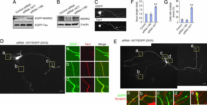
Fig. 1.
Suppressing MARK2 induces formation of multiple axons. (A) HEK-293 cells were transfected with the same amount of EGFP-MARK2 and tau together with control siRNA or MARK2 siRNA-1677 or -1180. Cell lysates were subjected to immunoblotting with antibodies against MARK2 or GFP. (B) Primary neurons were transfected with siRNAs and cultured for 72 h. The levels of endogenous MARK2 were assessed by immunoblotting and normalized with β-actin. (C) Hippocampal neurons transfected with EGFP plasmid were stained with Tau1 antibody at DIV3. (D) Neurons transfected with siRNA-1677/EGFP were stained with Tau1 antibody at DIV3. The boxed areas indicate dendrite-like (a) or axon-like (b and c) neurites. (E) Neurons transfected with MARK2-siRNA-1677/EGFP were stained with antibody against synapsin at DIV5. The boxed areas indicate dendrite-like (c and d) or axon-like (a, b, and e) neurites. (F) Quantitative analysis of axon length in siRNA-transfected neurons. All of the experiments were repeated at least three times. Data are shown as means ± SEM, and ≥60 cells were analyzed for each treatment per experiment. Axon length in control siRNA-transfected neurons was taken as 1.0. ∗∗, P < 0.01. (G) MARK2-siRNA-1677 increases the percentage of cells with multiple axons. Data are means ± SEM. ∗∗, P < 0.01.

